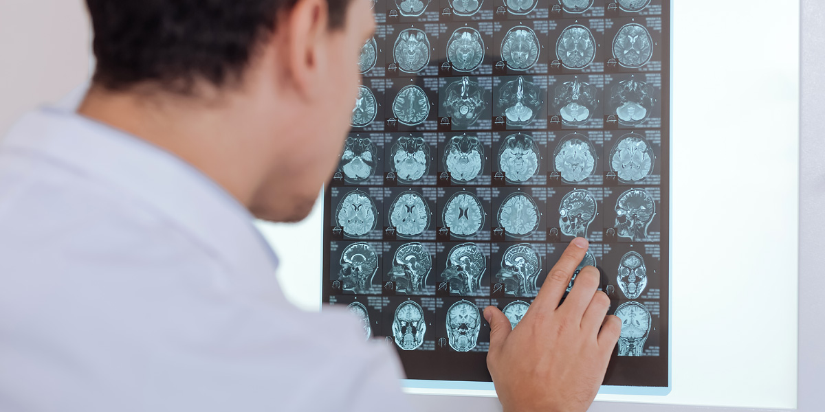
MRI, or magnetic resonance imaging, uses a magnetic field and pulses of radio wave energy to make pictures of organs and structures inside the body. During an MRI exam, the patient lies down on a moveable table that slides into the center of the magnet. Some magnets allow the patient’s head to remain outside of the magnet during the exam; others are open on all sides. The magnet causes the body’s protons to align themselves to receive radio signals. When radio waves are sent toward the lined-up atoms, they bounce back and a computer records the signal. Different types of tissues send back different signals.
The MRI at the MCH&HS Diagnostic Imaging Center features a large bore which creates a less stressful testing environment and more space for you to maneuver in an out of the machine before and after testing.
An MRI can be used to:
- Diagnose central nervous system disorders
- Identify brain tumors, strokes and chronic disorders of the nervous system
- Identify damage caused by heart attack or heart disease
- Detect blood vessel plaques and blockages
- Identify and diagnose bone and joint damage
- Reveal tumors and functional disorders in organs such as the lungs, liver, pancreas, kidney and spleen


