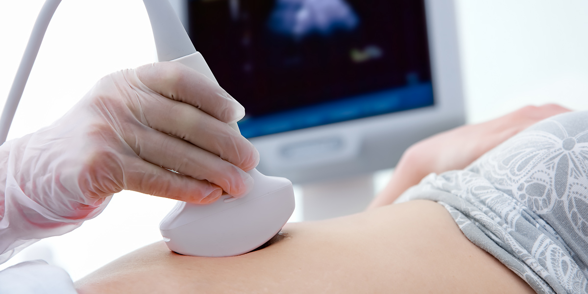
Ultrasound involves exposing part of the body to high-frequency sound waves to produce pictures of the inside of the body. Images are captured in real-time and show the structure and movement of the body’s internal organs, as well as blood flowing through blood vessels.
A transducer sends the sound waves and records the echoing waves. When the transducer is pressed against the skin, it directs small pulses of inaudible, high-frequency sound waves into the body. As the sound waves bounce off of internal organs, fluids and tissues, the sensitive microphone in the transducer records tiny changes in the sound’s pitch and direction. These signature waves are instantly measured and displayed by a computer, which in turn creates a real-time picture on the monitor.
Ultrasound is used to:
- Evaluate symptoms such as pain, swelling and infection
- Examine many of the body’s internal organs, including:
- heart and blood vessels
- liver
- gallbladder
- uterus, ovaries, and unborn child (fetus) in pregnant patients
- Guide procedures such as needle biopsies
- Image the breasts and guide biopsy of breast cancer
- Detect a variety of vascular conditions


